My research and investigation of hair loss
According to numerous studies worldwide, hair
loss has been proven a part of a clinically autoimmune disease, or human
immunity-associated disease, namely; human system as function protecting us
from dangerous diseases becomes a leading cause of hair loss. The negative response
to the pores can be seen with the microscopic images. The white blood cells of
the body have been affected including the pores. The concentration of abnormality
in follicles leads to a disruption of hair cells and stop growing, resulted in
the failure of tissue cells, the formation of T-cell, antibodies, and so on. As
hair loss occurs in the human defense mechanisms, hair cells stop growing (inactivates),
which can be seen clearly in the microscope images below; some hair roots are apparently
covered with white blood cell.

As shown in Figure, hair follicles are
surrounded by black spots; lymphocyte is a type of white blood cells. It’s
observed that those people who have been affected may have the smooth scalp from
hair follicles and the hair emerged from the skin is covered with thick
membrane. With the smooth scalp, this makes it difficult for conventional
treatment. Figure B and C shows unnoticeable hair follicles, and lastly, Figure
D represents the magnifying view of the systematic hair follicles.
Human immune surrounded stops growth. Figure
C skin is covered with a membrane, preventing the fair from the regeneration out
of the skin.

[Sebaceous Gland]
[Hair Follicles]

Figure shows the plugged follicles that hair
root cells cannot grow.

Figure shows surface surrounded by sebaceous
cell.
Figure shows hair is covered with connective
tissue membrane.


Both images above represent the follicles and
hair root cells of those who have been affected by hair loss, surrounded by
white blood cells and fat cells. They both are common in the similar point; the
magnification is to view clearly the white blood cells in which hair follicles
are swallowed; that resulted in the completely disability of the follicles. As time
has gone by, this becomes a thick membrane and treatment in the process is likely
to be ineffective. The figure below shows hair follicles with hair loss for
long time.


As shown in two images above – the membranes
(at the top and bottom of the follicles) are closed and somewhat thicker than
the previous image. The pores become prominent and the scalp has flat and thick
membrane physically, resulted that the patients have not been responsive to any
treatments in which thick membrane is laid.


Treatment – the drugs used are proven to be
ineffective compared to those untreated patients. At the initial phase of hair
loss, most patients are likely to be hopeful with hair growth without awareness
of sufficient condition, this is worse when time passes by.

Image of skin - Integumentary System – microscope
for skin surface, bright orange-red root, purple hair follicle surrounded,
thick hair within green connective tissue covered with purple epidermis along
the top edge of the 10X extension.
My research and investigation
Suspend antibodies and others in the
follicles, stop the accumulation of fat in the body caused by steroid hormones,
and get rid of the accumulated fat.
Health record (by the end year 2013)
- Weight: 67 kg.
- Length: 171 cm.
- Pressure 130/90 mmHg – intake of antihypertensives Atenolol 50 mg + Felodipine
(Plendil) 5 mg for more than 15 years
Health record (at present)
- Weight: 56kg. .
- Pressure 103/71 mmHg - normal blood
pressure, discontinuing antihypertensives
My profile of the experimental results
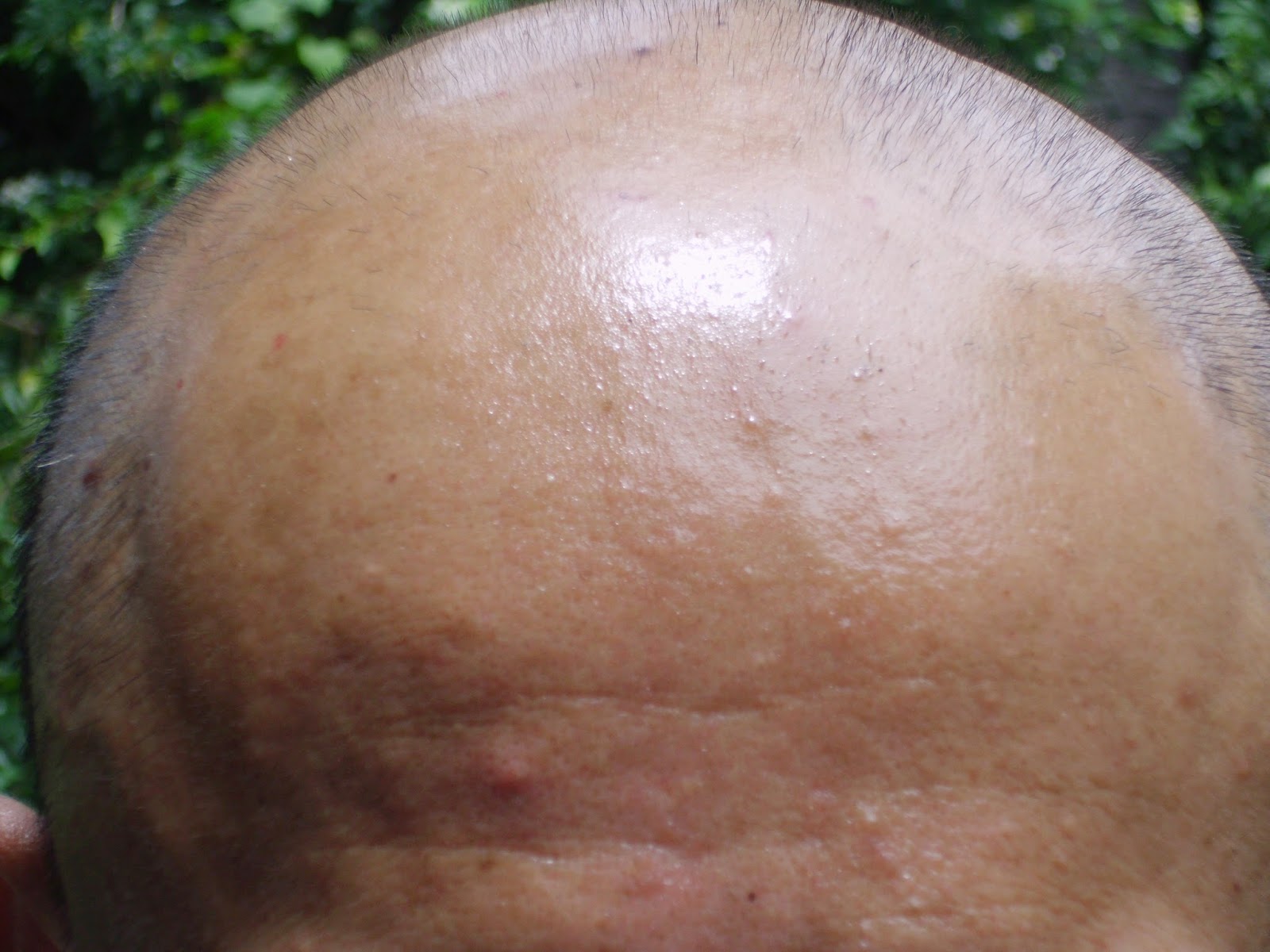
Photograph of the end of the previous year


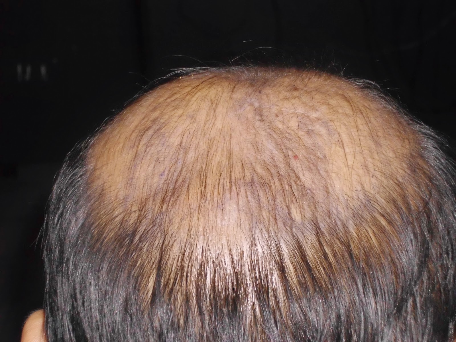
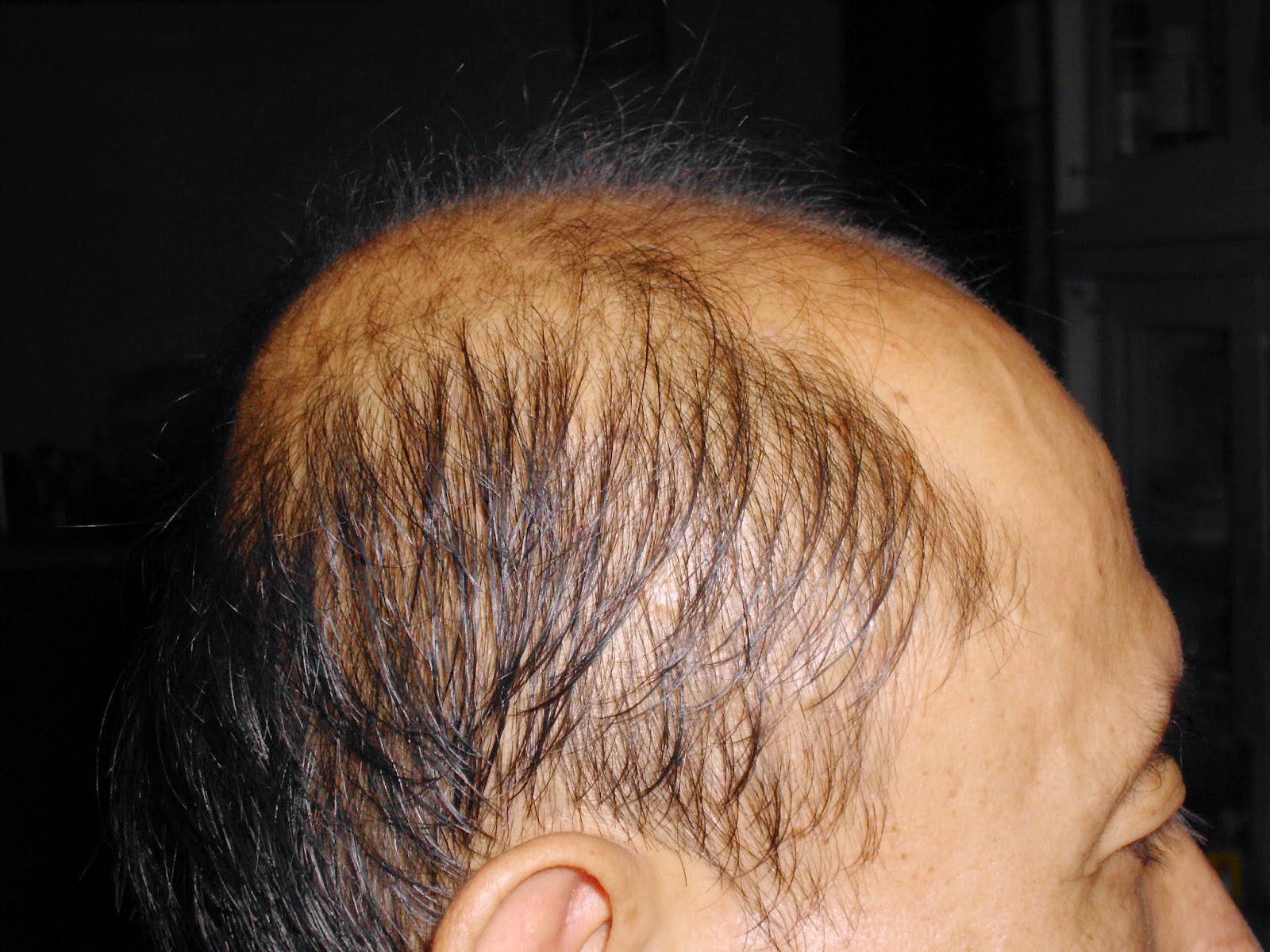
Photograph of the present year


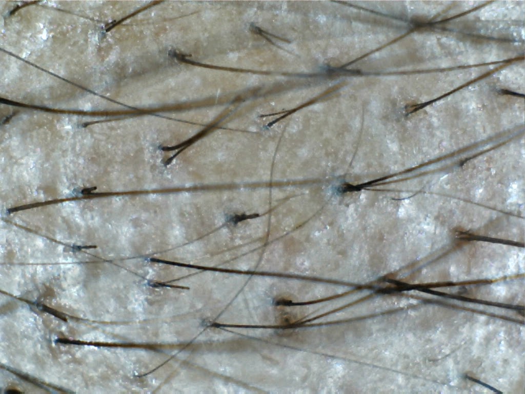
Photographs of microscope and experiment test


Video by microscope

This image shows stained scalp to distinguish
the disintegrated connective tissue membrane.

ความคิดเห็นนี้ถูกลบโดยผู้ดูแลระบบของบล็อก
ตอบลบ